|
Supplier |
accela s.r.o. |
|
Year of acquisition |
2018 |
|
Price |
14.6 million. CZK |
|
Financing |
OP VVV CORE FACILITIES CZ.02.1.01/0.0/0.0/16_017/0002515 |
|
Responsible Person |
doc. PharmDr. Martin Štěrba, Ph.D. |
In vivo experiments on experimental animals are still a key and indispensable part of medical research, because this approach, unlike in vitro experiments, reflects the complex environment of the whole living organism. Non-invasive imaging methods are an integral part of modern experimental research in this field as they can provide key anatomical and functional data.
Non-invasive ultrasound (US) imaging has had a firm place in clinical medicine for many decades. This approach has extraordinary translational potential because the same or similar parameters as in clinical practice can be evaluated in an experimental model. Other advantages of ultrasound include the ease of performing repeated examinations with minimal burden on the experimental animal and negligible operating costs. With ultrasound, the development of a pathological condition and its therapeutic influence (e.g. by a drug or surgical intervention) can be monitored in an experiment. Compared to invasive investigative approaches, this approach is therefore considerably more gentle on the experimental animal and can significantly reduce the number of experimental animals, which is in line with current trends and ethical requirements ('reduction' and 'refinement').
Most in vivo biomedical research today is carried out on small laboratory animals -usually rodents (especially mice and rats) and rabbits. However, the use of small laboratory animals places very high technical demands on the instrumentation. For example, the imaging capabilities for echocardiographic examination of mice must be prepared and optimized to ensure that the longitudinal dimension of the adult mouse heart is less than 1 cm and the transverse dimension is less than 0.5 cm. The physiological heart rate of the mouse is 450-650 beats/min. In addition, the examination must be performed under controlled anaesthesia with careful monitoring of vital signs.
In response to these specific requirements of experimental research, special high-frequency ultrasound equipment was developed for experiments on small laboratory animals only.
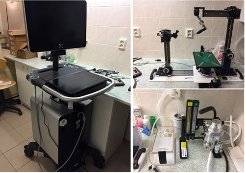
An example is the state-of-the-art VEVO 3100 high-frequency ultrasound set-up, complete with a full range of probes (18-70 MHz, with resolution up to 30 μm) for use on laboratory rodents (mouse, rat, guinea pig) and rabbit. The assembly forms a functional unit that allows a wide application of the instrument in experimental in vivo research. The assembly is designed for use by multiple research groups in a "Core Facility" format.
High-frequency UZ VEVO 3100 with LCD screen and touchscreen with relevant software and special software packages for individual applications + VevoLab software for off-line evaluation. Hardware and software are fully prepared for basic and more advanced ultrasound examinations (including 3D, ultrasound contrast, speckle tracking).
Full range of ultrasound probes: 1. MX201 18 MHz (10-21 MHz, max. resolution 100 μm, max. depth 4 cm, max. 367 FPS), 2. MX250 24 MHz (14-28 MHz, max. resolution 75 μm, max. depth 3 cm, max. depth 367 FPS), 3. MX250 24 MHz (14-28 MHz, max. 367 FPS), 3. MX400 38 MHz (21-44 MHz, max. resolution 50 μm, max. depth 2 cm, max. 449 FPS), 4. MX550D 50 MHz (26-52 MHz, max. resolution 40 μm, max. depth 1.5 cm, max. 557 FPS), and 5. MX700 70 MHz (30-70 MHz, max. resolution 30 μm, max. depth 1.2 cm, max. 476 FPS).
Vevo Imaging Station 2 – apparatus for fixing the animal and probe and moving both the animal and probe in all axes (for most examinations this is a necessity - examination directly "by hand" is very demanding and not very reproducible). Module for ultrasound guided injection.
Heated plate for mouse and rat with vital signs monitor (rectal temperature, ECG, heart and respiratory rate are monitored on-line, displayed on the LCD monitor of the ultrasound machine and stored together with the recording).
Inhalation anaesthetic unit for isoflurane anaesthesia with oxygen for breathing, including induction chamber and exhaust to absorber.

Examples of experimental research areas where the instrument can be used:
Cardiovascular research – basic and advanced examination of the structure and function of the heart (including e.g. myocardial strain examination) and blood vessels - applications in research: myocardial infarction, myocardial hypertrophy, heart failure, cardiotoxicity, cardiomyopathy, valvular defects, atherogenesis.
Tumors – orthotopic and s.c. implanted tumor imaging, evaluation of growth /3D volume, angiogenesis and tumor perfusion, controlled injection into the tumor.
Embryo-fetal applications – early detection/confirmation of pregnancy, teratogenicity studies of drugs and xenobiotics, fetal programming, ultrasound-guided puncture of amniotic fluid and left ventricle, intra-amniotic inflammation, placental structure and function, including assessment of maternal and fetal perfusion.
Liver disease – alcoholic and non-alcoholic fatty liver disease, hepatotoxicity, hepatoprotection and regeneration, changes in liver perfusion and liver metastasis.
Kidney disease – research on renal dysfunction, renal perfusion, chronic kidney disease, polycystic kidney disease, hypertension, hydronephrosis and renal cell carcinoma.
Galleries from the examination with this device are located on the manufacturer's website: https://www.visualsonics.com/product/imaging-systems/vevo-3100.
The manufacturer provides advanced hands-on training and instruction for individual applications.
Author:: doc. PharmDr. Martin Štěrba, Ph.D.