|
Supplier |
PE Systems s.r.o. |
|
Year of acquisition |
2018 |
|
Price |
12.7 milion. CZK |
|
Financing |
OP VVV CORE FACILITIES CZ.02.1.01/0.0/0.0/16_017/0002515 |
|
Responsible Person |
prof. MUDr. Stanislav Mičuda, Ph.D. |
Developing new drugs, discovering and synthesising new compounds that could help us fight cancer is the goal of many research teams around the world. This process is lengthy, challenging and can take many years. During this process, it is important to find out whether a newly discovered substance has an anti-cancer effect, whether it kills cancer cells or merely slows down their growth, etc. In the early stages of drug development, we test candidate substances in vitro, where we observe these processes on individual cells and try to unravel the mechanism of action at the molecular level. If at this stage the substance is shown to have promising properties, it can then be taken to the next step, which is to see how the organism as a whole reacts to the substance. In vivo testing in model organisms serves this purpose.
The priority for us is not only to find out how the substance will affect a healthy organism, or how large a quantity might be harmful, but more importantly how effective it will be specifically against cancerous growth. Since in model organisms (mice, rats,...) the occurrence of tumours is random and heterogeneous, we need to reach a state where we can test the substance reliably, i.e. to create "tumour models" where we can see the effect of the substance on the tumour cells we choose.
Comment:
Why do I need to use this procedure? There are many processes and processes in a living organism that influence and enable the organism to survive and adapt, whether to the environment or to emergencies. These processes may also influence the fate of the test substance in the organism. They may influence its absorption, metabolism, mechanisms of action or excretion.
Now we come to the question, how to non-invasively monitor, the effectiveness of the substance? That is, has the tumour stopped growing or has it shrunk, for example?
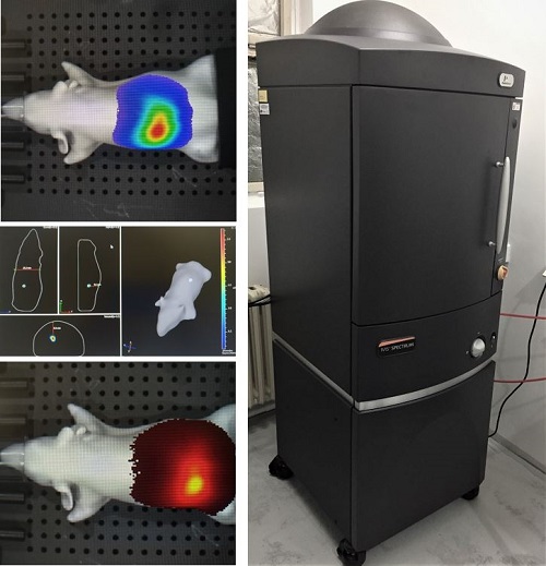
In the past (and sometimes even today), the size of subcutaneous tumours was (is) measured with a callipers, which is an instrument that can be used to determine the dimensions of the tumour and then calculate its volume. Modern medicine, however, has more modern means, based primarily on imaging techniques. These allow us to look inside the body without violating its integrity or intervening in any invasive way. Classical imaging techniques may include CT, MRI, X-ray or ultrasound, adapted to work with small laboratory animals. A more advanced option is to observe targeted structures within the body of the organism. Some devices use the principle of luminescence or fluorescence to image specific tissues or cells. By binding specific fluorescent or luminescent markers in the target structure of the organism/tumor cells and then binding these markers, these cells and structures can be imaged. This is made possible, for example, by the unique IVIS Spectrum In Vivo Imaging System from Perkin Elmer. The instrument detects both fluorescence (the emission of radiation induced by another excitation radiation and persisting only nanoseconds after the excitation has ended) and bioluminescence (the ability of organisms to produce light, e.g. enzymatically by luciferase to cleave D-luciferin to produce yellow-green light).
Key features and benefits of the IVIS Spectrum imager
High sensitivity in vivo fluorescence and bioluminescence imaging
Multi-object analysis (5 mice) with 23 cm field of view
High resolution (up to 20 microns) with a 3.9 cm field of view
Twenty-eight high-efficiency filters with a range of 430 - 850 nm
Ideal for resolution of multiple bioluminescent and fluorescent reporters (labeling)
3D diffusion tomographic reconstruction for both fluorescence and bioluminescence
Ability to import and automatically copy CT or MRI images with functional and anatomical context
Inhalation anaesthesia inlet and outlet ports

One of the interesting and useful functions of the instrument is the creation of a 3D spatial reconstruction of the observed object. This function works on the principle of creating a sequence of images, from which the instrument then composes a three-dimensional model, on which the exact location of the observed object (for example, a tumor) can be tracked, with possible measurement of signal intensity and dimensions. The locations of the individual organs from the available libraries can then be added to the three-dimensional models to complete the integrity of the observed model.
There are many possible applications of the instrument. It is possible to observe individually labeled cells in a plate/tube or whole live models, especially of laboratory mice of different species. By coupling a luminescent or fluorescent label to a specific antibody that can be directed to a variety of targets in the imaged model, we are then presented with a much wider range of possibilities in the observation of structures and tissues, whether healthy or pathologically altered, in a living organism. In this case, it should also be possible to observe certain ongoing infections. The instrument has great potential and the possibilities of its use will certainly grow in the future. At present, our department is the only one in the Czech Republic that has such a device.
Author of the text: RNDr. Martin Uher
Source:
IVIS Spectrum In Vivo Imaging System. PerkinElmer [online]. Waltham: PerkinElmer, 2019 [cited 2019-08-05].